Objectives
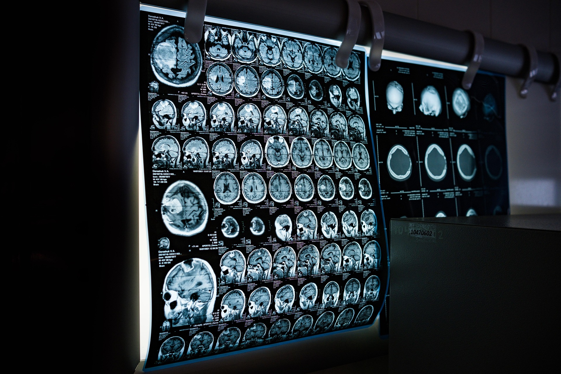
Starting from the physical principles of clinical and preclinical imaging, the students are introduced to various imaging modalities available at the Turku University Hospital and at the Turku Center for Disease Modelling. The course consists of lectures and Moodle-based assignments to give the student a basic understanding of imaging modalities used in the hospital and provide the knowledge basis for more advanced courses. On the completion of the course, the student will be able to:
- Explain the physical principles how different medical imaging modalities operate (X-ray, MRI, Nuclear Medicine)
- Understand the basic ideas of tomography for imaging (CT, PET)
- Give examples of the technical details for various medical imaging equipment
- Describe what is ionizing radiation, how we protect ourselves from it and how the use of it is controlled in the legislation
- Lärare
Jani Saunavaara
Objectives
Bioimage Informatics is a field of science that deals with the processing, analysis, visualization, handling and annotation of bioimages, using specialized software tools. An important aim in the field is to extract quantitative information from digital biomedical images. Starting from the fundamental concepts of digital images in biomedical sciences, students are introduced to various image processing and analysis techniques and tools. Along with lectures, demonstrations will be arranged to provide hands on training in image processing and in-depth and intuitive understanding of modern bioimage informatics approaches and applications. Further studies around the topic can be continued on course BioImage Informatics 2.
After the course, the student will be able to identify common phases in bioimage analysis, select proper methods and approaches for basic analysis tasks, explain the benefits of the use of bioimage informatics for quantification of biomedical images, and compare the differences of manual and computer based analysis of images.
- Lärare
Pekka Ruusuvuori
Common platform for Biomedical Imaging Programme students.
- Lärare
Riku Klén
- Lärare
Riku Klén
- Lärare
Päivi Koskinen
Objectives
The course will introduce students to the different seminar series held on the campus. After each seminar lecture the student will write a summary/abstract in the learning journal, focusing on the main message of the presentation. Upon completion of the course, the student is able to:
- Understand scientific presentation and communication in comprehensive way.
- Extract key scientific messages from the presentations and produce condensed summaries.
- Recall information and utilize note-taking during the presentation

- Lärare
Kari Vienola
Place: Teutori TEU103 "Sammio"
A practical course for any bioscientist or similar dealing with digital images, and needing to analyze their images, get numbers out of them, and evaluate when the analysis is correct.
On this course you will learn how to process and quantitatively analyze multi-dimensional digital bioimages and trouble-shoot problem situations. The focus is more on how to do things in practice, from the point of view of an image analyst or life scientist, and not so much from the point of view of a software developer.
Modes of study: The course consists of interactive physical sessions and related exercises. The course will be evaluated based on the submitted exercises and a final exam. Passing the course also requires participation in the sessions. Activity during sessions will give extra points.
- Lärare
Pasi Kankaanpää, Junel Solis
Objectives
By choosing a project topic of their choice, the students will familiarize themselves with a research field related to the biomedical field, ranging from medical imaging used by the University Hospital to image analysis on human-derived image data done by various research groups within the university. The goal is to apply their knowledge and gain deeper understanding into the use of these techniques, whether it is medical imaging or ex vivo image analysis, and how they can improve future patient diagnostics. On the completion of the course, the student will be able to:
- Explain and understand in detail the techniques used in the project (whether it is image analysis or image acquisition).
- Identify key elements for a standard project workflow.
- Design/Execute a project in a company or university setting.
- Lärare
Kari Vienola
Objectives
The aim is to provide listeners with information on the latest PET / TT / MRI imaging research topics and results as well as new technology developments in the field and topical issues both locally and internationally.
- Lärare
Riku Klén
The students are introduced to planning and performing a research project in the field of biomedical imaging as well as to critical, scientific thinking. Students will learn to plan and report the planning strategies of their research project, execute the experimental part/data collection, and orally present their research work.
The Master's thesis project will be conducted under supervision in a research group. Before starting the practical part, a student must submit a thesis information form and a thesis plan and present the plan in Thesis plan seminar. Responsible professor of the programme need to approve the thesis plan before the practical part can begin. The subject of the project need to be first discussed with the responsible professor. The project duration is approximately five months of intensive work with the practical part of the project. The results of the thesis project will be presented in the Master's thesis seminar New Discoveries.
Students are responsible for finding their own thesis projects.
The credits are part of the master's thesis for students in biomedical imaging and are given for the thesis plan, the thesis plan seminar, the thesis seminar and the practical laboratory work/data generation for the thesis.
The written thesis consists of the relevant review of literature, aims of the study, materials and methods, results and conclusions. Thesis will be accepted by the responsible professor of the programme and evaluated by two examiners.
- Lärare
Riku Klén, Kari Vienola